Volume 5, Issue 12
December 2025
Orthodontic-Related Infections: Diagnosis and Management of Infection Complications
Waleed Hashim Farran, Shatha Hamad Alharbi, Khadijah Mustafa Saidi, Fatima Abdullah Alhasawi, Eilaf Ahmed Alshahrani, Amjad Mohammad Alasmari, Hadeel Ibrahim Abughaniah, Fatima Mehar Aurangzeb, Marwa MohammedAmin Habhab, Almothana Ahmed Almulhim
DOI: http://dx.doi.org/10.52533/JOHS.2025.51213
Keywords: Orthodontic complications, fixed appliances, white spot lesions, root resorption, periodontal disease, pulpal changes, orthodontic treatment risks
Orthodontic treatment plays a crucial role in the correction of malocclusion and the enhancement of both dental aesthetics and functional occlusion. However, the implementation of such treatments frequently presents a range of potential complications and infections that can significantly jeopardize oral health. The application of fixed orthodontic appliances, in particular, creates a unique oral environment that favors the accumulation of dental plaque. This accumulation poses challenges for patients striving to maintain optimal oral hygiene, thereby elevating the risk of proliferating pathogenic microorganisms such as Aggregatibacter, Actinomycetemcomitans, and Tannerella forsythia. These microorganisms are notoriously associated with periodontal inflammation, which can lead to heightened susceptibility to gingivitis and periodontitis. Moreover, the challenges presented by orthodontic therapy can result in the decalcification of enamel, a phenomenon often evident as white spot lesions, largely due to the difficulties patients encounter when cleaning around brackets and wires. In addition to these issues, other significant complications may arise, such as root resorption, alterations in pulp health, and mucosal trauma. These factors may further predispose patients to various infections, painful ulcers, and opportunistic infections like candidiasis. In certain cases, the forces exerted during orthodontic treatment can exacerbate existing temporomandibular joint disorders, particularly in individuals with class II and III malocclusions or occlusal interferences. The magnitude of these adverse effects can be influenced by numerous factors, including the specific type and duration of orthodontic treatment, the intensity of applied forces, individual susceptibility, and baseline oral hygiene practices. To effectively mitigate these risks, it is critical to implement preventive strategies such as the utilization of fluoride-releasing bonding materials, regular fluoride applications, and resin infiltration techniques for the management of white spot lesions. Additionally, comprehensive patient education regarding rigorous oral hygiene practices is essential. Clinicians should also evaluate individual risk profiles, including systemic health conditions and unique oral anatomical considerations, before commencing orthodontic therapy. This review underscores the imperative of early diagnosis, meticulous microbial monitoring, and proactive management strategies to minimize the impact of complications and infections associated with orthodontic treatment, thereby promoting successful outcomes and enhanced patient satisfaction.
Introduction
Orthodontic treatment aims to correct malocclusion to improve dental and facial aesthetics and occlusal function (1). The first permanent molars are used as reference points in the dental arches. Orthodontic treatment aims to achieve normal occlusion using the first permanent molars (1). Bravo et al. (2) stated that there is a difference between normal occlusion and ideal occlusion. Ideal occlusion implies the hypothetical perfect occlusion, while normal occlusion can refer to a certain type of occlusion (1). Orthodontics is the science that covers the growth of the face and the jaws by changing the position of the teeth. It studies the factors, whether internal or external, that affect the development of the face and jaws (1, 3). Malocclusion is the third most common dental problem. For instance, in Asia, the percentage of class II and class III angle malocclusion is 21.42% and 5.76%, respectively (1, 3). Malocclusion negatively affects facial aesthetics. Treating malocclusion improves the patient's function, psychological status, and aesthetics (3). These negative effects impact women more than men. Hence, women seek orthodontic treatment more than men (3, 4). However, orthodontic treatment has negative effects, such as periodontitis, root resorption, pulpal damage, allergic reaction, and enamel demineralization (3). Post-orthodontic treatment, patients experience improved aesthetics, function, mastication efficiency, and reduced speech problems, consequently improving quality of life, self-confidence, and psychological status (3, 5). Adolescents and children who receive orthodontic treatment experience improvements in their lives, specifically in their social and emotional status (6). Children and adolescents have a significant overjet, which makes them more prone to traumatic dental injuries (7). If children receive interceptive orthodontic treatment, traumatic dental injuries decrease significantly, especially in children between 10 and 12 years of age (3). Additionally, myofascial pain and muscle tenderness decrease significantly post-orthodontic treatment (8). The perception of parents towards orthodontic treatment. Lack of parental awareness towards malocclusion and its effect on children's quality of life. Additionally, parents worry that their children will suffer dietary and speech problems (3). Education and the socio-economic status of the parents play a crucial role in the parents' perception towards orthodontic treatment (3). However, orthodontic treatment has some adverse effects on the teeth as well. It damages the crown of the teeth on which the brackets were put. This damage occurs through decalcification, fracture, erosion, decay, and discoloration (3). Fixed orthodontic appliances cause several adverse effects in the oral cavity. For instance, it increases the attachment of debris, plaque, and calculus (3). It extends the stagnation areas and makes maintaining oral hygiene difficult. Hence, increases the risk of tooth caries (3). Therefore, it is essential to weigh the pros and cons of orthodontic treatment to avoid unwanted results and obtain the most beneficial results (9). This review article seeks to comprehensively explore the various potential complications and infections associated with orthodontic treatment. It aims to provide insights into the diagnostic methods for identifying these issues, as well as effective management strategies to mitigate their impact on patient outcomes and overall treatment success.
Methodology
This narrative review is based on a comprehensive literature search conducted on May 22, 2025, using ScienceDirect, PubMed, Wiley Library, Dynamed, MDPI, Oxford Academic, BMC, and Cochrane databases. The research utilized Medical Subject Headings (MeSH) terms and relevant keywords, such as orthodontic treatment and its effect on the oral cavity, to identify studies that examined orthodontic treatment and its impact on the changes in the oral cavity. A manual search was also conducted using Google Scholar, and the reference lists of identified papers were reviewed to locate additional relevant studies. No restrictions were applied regarding publication date, language, participant age, or type of publication, ensuring a broad exploration of the available literature.
Discussion
Orthodontic-Related Infections
Oral hygiene is compromised during orthodontic treatment, especially when fixed orthodontic appliances are installed. Decreased oral hygiene leads to the development of caries and gingivitis, which in turn leads to loss of gingival attachment (10). Additionally, the presence of orthodontic appliances increases the stimulated salivary flow rate, the buffer capacity, the PH, and the plaque index scores (10). Additionally, it was found that the levels of Lactobacilli increased after three months of initiation of orthodontic treatment. Moreover, the oral environment changes significantly after the placement of fixed orthodontic appliances. For instance, pocket depth, bleeding index, and plaque index increased significantly (10). After six months of fixed orthodontic appliance placement, oral microflora shifted to disease-inducing bacteria, in addition to the increase of spirochetes and fusiform bacilli (10). Orthodontic treatment is not the cause of dental caries and gingivitis. However, it is a cause of exaggerated inflammation or caries due to poor oral hygiene (10). Orthodontic appliance placement poses a challenge to maintaining optimal oral hygiene. Additionally, the increase in bacterial count in the saliva, especially Prevotella intermedia and Lactobacilli, renders the gingival tissues and teeth more susceptible to developing inflammation and caries, respectively (10). However, good oral hygiene does not guarantee decreasing plaque index, since teeth crowding makes it difficult to control plaque and efficiently remove it (10). This is attributed to the increasing plaque-retentive areas and the inaccessibility to these areas while having a fixed orthodontic appliance (10, 11).
Periodontal diseases include gingivitis, periodontitis, and loss of periodontal support (9). The bacteria present in the accumulated dental plaque can cause periodontitis, gingivitis, and loss of alveolar bone during orthodontic treatment (9). Plaque is a crucial factor in developing several dental problems; however, it is not the only factor that contributes to developing such diseases. For instance, the soft tissue reaction to metallic brackets, the presence and composition of calculus, the patient’s immunity, and the presence of systemic diseases, such as uncontrolled diabetes, play a role in the development of gingivitis and periodontitis (12, 13). Moreover, metallic brackets can cause localized gingival inflammation and change the bacterial oral flora (9).
Orthodontic treatment has negative effects on supporting alveolar bone and roots. For instance, dehiscence in the upper and lower canines occurs due to the placement of fixed orthodontics and the force applied to them. An adequate amount of healthy gingival tissues is required to withstand orthodontic treatment without leading to gingival recession or dehiscence (14). For example, when labial bodily movement was applied to the lower incisors in the presence of gingival inflammation, gingival recession occurred (14).
Root resorption is a pathological process in permanent teeth. Root resorption has four grades, which are grade 0, characterized by no visible root resorption, grade 1, characterized by diffuse, slow, and mild root resorption, grade 2, characterized by moderate resorption, the apex starts to disappear or become semicircular and about 25% of the root is resorbed, whereas grade 3 is characterized by severe resorption where more than quarter of the root is resorbed (15). During orthodontic treatment, different grades of root resorption can occur, which can affect the health and integrity of the teeth. However, most root resorptions resulting from orthodontic treatment are insignificant and account for only 1 mm (11, 16, 17). The force applied to the teeth during the orthodontic treatment, the type of appliances used, the distance that the teeth move, and the duration of the orthodontic treatment can affect the health of the roots and rate of tooth resorption significantly (18). Additionally, root resorption incidence is higher in cases where extraction is done. Upper anterior teeth are more prone to root resorption due to factors such as thin roots and dilacerated roots (18). Vertical bone resorption can result from orthodontic treatment as well and cause loss of periodontal attachment (17). The exact cause of root resorption during orthodontic treatment and its degree are still unknown. However, there are some predisposing factors, such as root morphology, vitality of the teeth, Age, systemic diseases, genetic factors, patients' habits, and history of trauma to teeth (9).
Orthodontic treatment can cause temporomandibular joint dysfunction, which includes difficulty eating, pain, clicking of the jaws, and difficulty opening the jaws (19). Multiple factors play a role in the association between temporomandibular joint dysfunction and orthodontic treatment, such as class II and III malocclusion, occlusal interferences, significant overjet, or posterior crossbite, anterior open bite, functional appliances, intermaxillary elastics, and extraoral forces (20). However, orthodontic treatment can alleviate temporomandibular joint dysfunction when treating occlusal problems (20).
Metallic orthodontic appliances are widely used in orthodontic treatment; however, they are associated with a significant adverse effect: the decalcification of teeth. This condition is frequently indicated by the development of white spot lesions (Figure 1), which signify the early stages of enamel demineralization. Research indicates that approximately 50% of patients undergoing treatment with fixed metallic orthodontic devices may experience this issue. Notably, the upper anterior teeth are typically the most susceptible to the formation of white spot lesions. Remarkably, signs of decalcification can manifest as early as one month after the initiation of orthodontic treatment (21). Metallic orthodontic appliances can significantly contribute to the accumulation of dental plaque due to their intricate design, which complicates effective oral hygiene practices. This accumulation, coupled with alterations in the oral microflora, can result in a higher risk of enamel demineralization, ultimately compromising dental health (14). When the orthodontic brackets are removed, these spots decrease in size (14). When orthodontists observe the presence of white spot lesions following the removal of brackets, it is advisable for them to refrain from immediately recommending interventions such as fluoride application or microabrasion. Instead, these professionals should focus on closely monitoring the patient’s condition while allowing time for natural enamel remineralization to occur. In conjunction with this strategy, patients ought to be instructed on the importance of maintaining rigorous oral hygiene practices. Furthermore, the use of fluoride-containing mouthwashes can be beneficial in supporting the remineralization process and protecting enamel health (14).
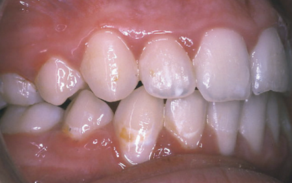
Figure 1: White spot lesion in the lower canine after orthodontic treatment (22).
Orthodontic treatment has a significant effect on the vitality of the teeth. For instance, rapid orthodontic movement of the teeth can result in pulpal injury. This is attributed to the alteration in the blood vessels in the periodontal tissues in the periapical area, and the blood vessels entering the pulp. Additionally, teeth may develop altered sensation to stimuli (23). Pulpal changes resulting from orthodontic treatment can have a direct effect on odontoblasts in completely formed teeth and Hertwig’s sheath in incompletely formed teeth. The severity of pulpal changes is directly proportional to the magnitude of orthodontic forces (23). Orthodontic forces, particularly those exerted in a labiolingual direction, induce tilting of the apical third of the teeth. This specific movement can lead to compromised collateral circulation, which ultimately results in pulpal degeneration. In adolescent patients, this phenomenon may manifest as the degeneration of some odontoblasts while others experience atrophy. To mitigate potential damage and facilitate the healing of dental tissues, it is advisable to apply intermittent orthodontic forces rather than continuous pressure, thereby promoting better tissue response and overall dental health (23). The respiratory rate within the dental pulp inversely correlates with the application of orthodontic force. During orthodontic treatment, a mean respiratory depression of approximately 27.4% is observed. As the force increases, the respiratory rate decreases, thereby influencing the maturation processes of the tooth (23). However, Consolaro et al. (24) indicated that the dental pulp exhibits a minimal reaction to orthodontic movements, typically characterized by temporary inflammatory responses. They further emphasized the importance of ruling out any history of trauma in cases where orthodontists encounter sudden pulp necrosis. If no such traumatic event is identified, practitioners must consider discontinuing the orthodontic treatment to prevent further complications (24).
The oral cavity is covered with a thin epithelial mucosal membrane. This membrane is easily ulcerated by sharp tooth edges, traumatic biting, and metallic orthodontic appliances (25-27). When the oral mucosa is ulcerated, it becomes easily infected by bacteria in the oral flora (25, 26). Traumatic ulcers are common findings in patients with fixed or removable orthodontic appliances. The direct contact between the soft tissues and the fixed appliances results in soft tissue ulcers. These ulcers are often seen in the tongue, buccal, labial, or lingual mucosa. Additionally, traumatic keratosis results from friction between the fixed orthodontic appliances and cheeks (9). Symptoms of traumatic ulcers are often burning pain. Orthodontists should put soft wax on the sharp edges of any appliance or modify it (9). If a patient with traumatic ulcers has poor oral hygiene, there is a risk of developing candidal infection (9). About 60% to 80% of oral ulcers that occur in orthodontic patients are traumatic, while 8% to 30% of the ulcers are aphthous ulcers (Figure 2) (28).
Papageorgiou et al. (30) conducted a controlled clinical investigation to assess the effects of fixed orthodontic appliances on the composition of the subgingival microbiota and the associated risk of periodontal infection. Collectively, these studies indicate that the placement of fixed orthodontic appliances induces a significant qualitative alteration in the subgingival bacterial consortium. Patients undergoing orthodontic treatment exhibit a markedly increased prevalence of Aggregatibacter and Actinobacillus actinomycetemcomitans within the gingival sulcus following the placement of these appliances compared to untreated controls. Notably, while the relative abundance of A. actinomycetemcomitans demonstrates a gradual decline in the months immediately following appliance removal, it remains significantly elevated relative to baseline levels observed in non-orthodontic cohorts. Furthermore, the study reported a sustained increase in detection rates of Tannerella forsythia for up to three months post-appliance removal. These microbial alterations likely reflect an overall increase in the intraoral microbial burden and a parallel inclination toward periodontal inflammation. This underscores the necessity for rigorous microbial monitoring and the implementation of adjunctive infection-control strategies throughout the duration of orthodontic therapy (30).

Figure 2: Aphthous ulcer in the labial mucosa (29).
Prevention of such complications is essential to mitigate their effects on the overall oral health during orthodontic treatment. Fluoride plays a pivotal role in the prevention of dental caries, particularly in orthodontic patients who are at increased risk due to plaque accumulation around fixed appliances. The utilization of fluoride-releasing bonding materials has emerged as a viable strategy for enhancing caries resistance during orthodontic therapy. Evidence suggests that the application of topical fluoride around orthodontic brackets at six-week intervals can significantly reduce the incidence of white spot lesions, an early sign of enamel demineralization. Furthermore, the routine incorporation of fluoride into orthodontic care protocols mitigates the cariogenic challenges posed by fixed appliances. It is crucial to note that successful orthodontic outcomes are closely linked to the patient’s baseline oral hygiene and caries risk; therefore, individuals must demonstrate adequate oral cleanliness and low caries activity before commencing treatment. In cases where white spot lesions develop, resin infiltration offers a minimally invasive option for aesthetic management post-debonding, although timely intervention is essential for optimal results (3).
Conclusion
Orthodontic treatment provides a wide range of aesthetic and functional advantages; however, it is also associated with several potential complications. Among the most common issues are periodontal infections, enamel decalcification, root resorption, and pulpal damage. To mitigate these risks, effective oral hygiene practices and preventive strategies, such as the application of fluoride, are essential. Clinicians should adopt a personalized approach to treatment, taking into consideration individual risk factors and the specific oral health status of each patient. Furthermore, patient education and regular monitoring are critical in preventing adverse outcomes. Striking an appropriate balance between the benefits of orthodontic intervention and the potential for complications is fundamental to achieving optimal long-term results.
Disclosure
Conflict of interest
There is no conflict of interest.
Funding
No funding.
Ethical consideration
Non applicable.
Data availability
Data that support the findings of this study are embedded within the manuscript.
Author contribution
All authors contributed to conceptualizing, data drafting, collection and final writing of the manuscript.
