Volume 5, Issue 11
November 2025
Effects of Chronic Illnesses and Medications on Pediatric Dental Development and Morphology
Wejdan Alharith, Noor Alsaffar, Ahmed Alotaibi, Ibtihal Algarni, Anfal AlmohammadSaleh
DOI: http://dx.doi.org/10.52533/JOHS.2025.51103
Keywords: pediatric dental development, chronic disorders, medications, cardiovascular diseases, endocrine disorders
Human beings are diphyodont, possessing primary and permanent dentition. The development of the primary dentition starts during the embryonic phase. The functional tooth germs initiate from the dental lamina of primary teeth in the maxilla and the mandible at six weeks in utero and start their mineralization at 14 weeks in utero. Whereas the formation of permanent dentition commences at 4 months in utero from the successional dental lamina that is lingually attached to the enamel organ of the primary teeth. The formation of primary teeth takes about 3.5 years from the eruption of the first lower deciduous central incisor till the completion of the roots of the second deciduous molars. Tooth buds for primary and permanent dentitions are vulnerable to systemic disorders, significantly affecting enamel and dentin formation. Enamel hypoplasia and other defects can arise from genetic conditions, salivary composition alterations, systemic diseases, or environmental factors. Systemic diseases that affect dental development are divided into primary and secondary disorders. secondary disorders result from events such as hormonal changes or trauma. Additionally, medications can significantly impact dental morphology. The prolonged use of sedatives and anticonvulsants can cause malocclusion, whereas corticosteroids can weaken the hardness of dental tissues, subsequently affecting mastication. Moreover, immunosuppressive medications are associated with abnormal development of craniofacial structures. Chronic diseases, such as cardiovascular disease, can negatively impact pediatric dental development. Children with congenital heart disease have shown poorer oral health conditions, increased carious lesions, and delayed dental development compared to healthy children. Endocrine disorders, such as hypothyroidism and growth hormone deficiency, also significantly affect craniofacial structures, resulting in complications such as delayed tooth eruption and enamel hypoplasia. This review article aims to underscore the relationship between chronic illnesses and medications and pediatric dental development, highlighting gaps in the existing literature, discussing such challenges.
Introduction
Human beings are diphyodont, which is defined as having primary dentition and permanent dentition (1). The development of the primary dentition starts during the embryonic phase. The functional tooth germs initiate from the dental lamina of primary teeth in the maxilla and the mandible at six weeks in utero and start their mineralization at 14 weeks in utero (2). Whereas the formation of permanent dentition commences at 4 months in utero from the successional dental lamina that is lingually attached to the enamel organ of the primary teeth (3). The formation of primary teeth takes about 3.5 years from the eruption of the first lower deciduous central incisor till the completion of the roots of the second deciduous molars (4). While the formation of permanent incisors, canines, and premolars starts at birth and takes about 13.5 years till the completion of the roots of the second premolars. Whereas the second and third molars' complete formation takes about 21 years (1). The first permanent molar is different from other molars; it starts formation at 4 months in utero, and its roots are completed around the age of nine (1).
The tooth bud, whether permanent or primary, is extremely sensitive to systemic disturbances, especially the enamel. Enamel and dentin defects result from alterations in genetics, the composition of saliva, an immunological response to cariogenic bacteria, systemic diseases, or environmental and behavioral factors (5, 6). These defects can be generalized in the whole dentition or localized in certain teeth. It can be symmetrical and cross the midline or asymmetrical. Additionally, the defects can affect either one surface of the tooth or involve more than one surface. Systemic diseases that affect the formation of the tooth or result in hard tissue defects are classified into primary and secondary systemic diseases (5). Primary diseases are defined as the disorders that originate from or affect several organs at the onset of the disease. These primary disorders include genetic, hereditary, and developmental diseases. Secondary disorders can be stimulated by an event such as changes in hormone levels, trauma, the onset of a disease, or viral infections (5).
Medications can affect the development and morphology of teeth as well. For instance, the long-term use of certain sedatives and anticonvulsants was found to be associated with changes in the oral and facial structure (7). Such changes can cause malocclusion, rendering orthodontic intervention a must. Additionally, the prolonged use of corticosteroids in chronic conditions such as asthma can cause changes in the hardness of dental or oral tissues, which negatively affect the child’s mastication and health. Immunosuppressants can negatively affect the development of the oral and facial bones as well (7).
This review article aims to elucidate the significant effect of chronic conditions and medications on the development and morphology of the dentitions of pediatric patients, aiming to highlight the gap in the existing literature.
Methodology
This narrative review is based on a comprehensive literature search conducted on September 1, 2025, using ScienceDirect, PubMed, Wiley Library, Dynamed, MDPI, Oxford Academic, BMC, and Cochrane databases. The research utilized Medical Subject Headings (MeSH) terms and relevant keywords, such as chronic diseases and their impact on the dental development of pediatric patients, to identify studies that examined the effect of chronic conditions and medications on the development of dentition of children. A manual search was also conducted using Google Scholar, and the reference lists of identified papers were reviewed to locate additional relevant studies. No restrictions were applied regarding publication date, language, participant age, or type of publication, ensuring a broad exploration of the available literature.
Discussion
Effect of Chronic Illnesses
Several chronic diseases affect dental health and dental development in pediatric patients, such as congenital heart disease. Congenital heart disease is defined as a morphologic malformation of the heart and intrathoracic great vessels that occurs during intrauterine development (8). It accounts for 0.5–0.8% of all live births (9). Recent advances in imaging modalities and diagnostic methods have significantly increased the survival rate of children with congenital heart diseases (10). However, the quality of life of these patients is compromised due to the presence of several oral health conditions resulting from congenital heart disease. Congenital heart disease leads to the development of carious lesions, dental plaque, and periodontal diseases (8). Additionally, it can cause severe growth retardation, especially in patients with right-to-left shunting, pulmonary hypertension, prominent cardiac insufficiency, and cyanosis (8). Saraç et al. (8) conducted a cross-sectional study on children with congenital heart disease and healthy children to assess the impact of the disease on their oral health. Although the difference between the two groups regarding the caries index was not statistically significant, children with congenital heart disease had a lower dental age despite having the same chronological age as the healthy group (8).
Endocrine disorders significantly affect the development of the body and can negatively affect the craniofacial structure (11). For instance, hypothyroidism can cause neurological, cardiovascular, gastrointestinal, and metabolic dysfunction. However, it severely affects the craniofacial structure, causing an anterior open bite, delayed tooth eruption, tooth impaction, retrognathism, micrognathism, and enamel hypoplasia (12). Additionally, growth hormone deficiency can cause an immature facial appearance, maxillary hypoplasia, anterior open bite, mandibular retrognathism, short mandibular ramus, and decreased anterior facial height (13). Obesity affects the integrity of craniofacial structure by inducing prognathism, early tooth eruption, periodontitis, and increasing facial height (14). Alfaro et al. (11) conducted a cross-sectional study on 1508 children and adolescents between 1 and 19 years, who had hypothyroidism, obesity, and non-syndromic hypogrowth. They found that children with medicated hypothyroidism had altered craniofacial structure in the sagittal, transverse, and vertical planes compared to healthy children (11). Additionally, they showed increased head circumference, zygomatic width, nasal base width, intercommissural distance, cranial base length, and maxillary and mandibular length (11). However, Vucic et al. (12) found that children with untreated hypothyroidism had decreased maxillary and mandibular growth, which could result in teeth impaction or delayed tooth eruption (12).
Diabetes mellitus type I occurs due to environmental, immunological, and genetic factors that destroy the pancreatic beta cells, which directly affects the production of insulin, causing hypoglycemia and hampers mineral homeostasis (15). Diabetes mellitus type I often occurs at the age of development of the primary and permanent dentition (16). Impeding mineral homeostasis directly affects the development of teeth (17). Pediatric patients with type I diabetes mellitus were found to have decreased vertical height of the alveolar bone and decreased cephalometric measurements. Additionally, mineralization and the ameloblast’s function are significantly affected due to the absence of secretory proteins, causing enamel and dentin defects (15, 18).
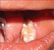
Yamunadevi et al. (15) conducted an observational study on 30 diabetic patients and compared them with healthy participants and found that 7 patients had a 6th cusp in the mandibular first molars, one patient had 5 cusps in the mandibular second molar, and one patient had 6 cusps in the second mandibular molars (Figure 1), one patient had a Y pattern in the mandibular first premolars, one patient had 4 cusps in the mandibular second premolars, one patient had flower-shaped mandibular molar (Figure 2), one patient had a prominent cusp of carabelli and oblique ridge in maxillary molar, 4 patients had microdontia, and 11 patients suffered attrition. Yamunadevi et al. (15) reported that type I diabetic patients showed morphological changes, especially in the number and shape of the cusps. The 6th cusp in the mandibular molars is classified into tuberculum intermedium and tuberculum sextum (19). If the extra cusp is present lingually is named tuberculum intermedium, whereas if it is located on the distal margin is called tuberculum sextum (19). Enamel hypoplasia in patients with diabetes mellitus can be attributed to the secondary complications associated with the disease, notably microangiopathy (20). This condition impairs the integration of calcium into the enamel matrix while simultaneously affecting the proteins responsible for regulating the precise arrangement of enamel prisms (20). Consequently, the ameloblasts' function is significantly diminished. Additionally, elevated blood glucose levels directly disrupt the structural integrity of the enamel prisms by affecting the activity of secretory ameloblasts (20).

Figure 1: Left mandibular first molar with 6 cusps and second molar (15).
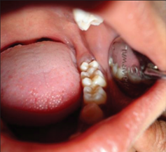
Figure 2: Flower-shaped left permanent mandibular first molar (15).
Chronic kidney disease is the presence of an injury or loss in renal function for three or more months. Although chronic kidney disease is not common in children, it often has a devastating effect on their health (21). However, the incidence of chronic kidney disease is growing among children, accounting for 85 cases per million age-related population (pmarp) in the USA and 62 pmarp cases in Europe (21). Chronic kidney disease affects bone and mineral metabolism, which can contribute to maxillofacial manifestations, including both soft and hard tissue changes (22). The various oral manifestations of chronic kidney disease are observed in about 90% of patients (23). For instance, chronic kidney disease affects the maxilla, mandible, and alveolar bone metabolism and alters the turnover of the maxillary cortical bone. Additionally, it causes gingivitis, periodontitis, gingival enlargement, halitosis, leukoplakia, pallor of the oral mucosa, dysgeusia, xerostomia, and candidiasis (22, 24). Renal osteodystrophy and hyperparathyroidism associated with chronic kidney disease have serious effects on the development of the teeth and jaws, such as jaw enlargement, bone demineralization, delayed tooth eruption, radiolucent giant cell lesions, dental spacing, tooth mobility, decreased bone trabeculation, radiolucent fibrocystic lesions, propensity for mandibular and maxillary fractures, abnormal bone healing after tooth extraction, and hypercementosis (22, 25).
Despite the several manifestations of chronic kidney disease on the maxillofacial structure, developmental defects in enamel are the most significant and prevalent manifestation among pediatric patients. Developmental enamel defects are disturbances in the enamel matrices and the mineralization of enamel during the odontogenesis process. This process occurs at approximately 3–4 months of intrauterine life for deciduous teeth and from birth for permanent teeth (22). Chronic kidney disease causes developmental defects in enamel due to hypocalcemia, decreased serum levels of 1,25-dihydroxycholecalciferol, and elevated levels of serum phosphate, the parathyroid hormone, and fluoride, which negatively impact the amelogenesis process (22). Nunn et al. (26) conducted a prospective study and found that approximately 83% of the patients had diffuse opacity, and 22% of them had enamel hypoplasia. Whereas Al Nowaiser et al. (27) reported that 57% of the participants had developmental enamel defects.
Effect of Certain Medications
Medications can affect the dental development in pediatric patients. For instance, tetracycline antibiotics are a recognized cause of intrinsic teeth discoloration. Tetracycline chelates with the calcium ions found during tooth development, resulting in the formation of tetracycline-calcium orthophosphate complex in teeth (28). When this complex is exposed to light, it oxidizes, resulting in intrinsic tooth discoloration. The intrinsic discoloration is often observed in the cervical part of the tooth. Additionally, the appearance of the discoloration can be yellow, grey, or brown. The intensity of discoloration depends mainly on the dose and period of administration (28). Therefore, tetracycline is avoided during the second and third trimesters and in children up to the age of eight. Minocycline, which is a derivative of tetracycline, causes discoloration of fully erupted teeth in addition to discoloration of the gingival tissues and alveolar bone (29). The minocycline stains have a blue-grey hue and often appear in the incisal and middle thirds of the tooth. If the stains extend to the roots, it causes a black or green discoloration (28). This discoloration was noted after the extraction of these teeth. Additionally, minocycline causes alveolar bone pigmentation, which can be clinically observed beneath the semitranslucent maxillary and mandibular anterior alveolar mucosa, the mandibular posterior lingual mucosa, and the hard palate (28). The incidence of minocycline-induced bone pigmentation is approximately 10% after one year in patients taking at least 100 mg of minocycline daily, while the incidence increases to 20% after four years, which is irreversible (28). Doxycycline has also been associated with the discoloration of the erupted teeth. Whereas ciprofloxacin is associated with green intrinsic discoloration if used in infants (28).
Antiresorptive medications that are administered during the neonatal stage or childhood are strongly associated with dental defects. Antiresorptive medications, such as bisphosphonates, inhibit osteoclastic bone resorption, which is used for the treatment of skeletal disorders, including osteoporosis, Paget’s disease, osteogenesis imperfecta, and metastatic bone diseases resulting from pathological bone resorption (30). Most pediatric patients who receive antiresorptive medications for the treatment of osteogenesis imperfecta start medication at the age of four, with some initiating the treatment at two weeks old. Bisphosphonates delay tooth eruption, increase the number of impacted teeth in patients with dental disorders, such as agenesis, apically extended pulp chambers, dens invaginata, and denticle (30). Osteogenesis imperfecta patients who receive bisphosphonates have lower dental age, delayed dental maturity, and tooth eruption if they were administered it before two years of age (30). Additionally, they have an increased risk of experiencing abnormalities in tooth formation, displaying as morphological aberrations, tooth agenesis, and enamel defects (30). However, some studies reported that the prevalence of missing or unerupted teeth in osteogenesis imperfecta patients varies according to the nature of the disease, regardless of receiving bisphosphonates (30). Therefore, bisphosphonates have been associated with delaying tooth eruption in patients with osteogenesis imperfecta; however, antiresorptive medications were not associated with delaying tooth eruption in other diseases (30).
Conclusion
Chronic diseases and medications that occur during childhood can significantly affect the dental development of pediatric patients. Cardiovascular diseases, chronic kidney diseases, diabetes mellitus, and medication, including antibiotics, such as tetracycline and minocycline, and antiresorptive medications, such as bisphosphonates, can have a pronounced effect on the dental development of pediatric patients. These effects include delayed eruption, morphological changes, and intrinsic discoloration of the teeth. Such effects can facilitate early diagnosis of some diseases, and others can be reversed if detected early. Further guidelines and clinical recommendations are required to facilitate the early detection of these developmental defects, in addition to taking precautions when certain medications are administered.
Disclosure
Conflict of interest
There is no conflict of interest.
Funding
No funding.
Ethical consideration
Non applicable.
Data availability
Data that support the findings of this study are embedded within the manuscript.
Author contribution
All authors contributed to conceptualizing, data drafting, collection and final writing of the manuscript.