Volume 5, Issue 11
November 2025
Antineoplastic Drugs and Their Effects on Soft Tissue and Bone Remodeling in the Oral Cavity
Mohammed Abdullah Batwa, Reem Fahad Alsuwaidan, Mohammed Ali Alzahrani, Mohammed Ghazi Aljohani, Hala Atif Albalawi, Mohammed Salem Alnhhale, Talal Ghazi Almarwani, Aliya Salem Alqhatani
DOI: http://dx.doi.org/10.52533/JOHS.2025.51116
Keywords: antineoplastic drugs, oral cavity, adverse effects, mucosa, bone
Antineoplastic drugs, commonly employed in cancer treatment, have notable adverse effects on soft tissues and bone, often resulting in serious complications. These agents, which include those targeting DNA synthesis, mitotic inhibitors, and DNA-interactive compounds, disrupt tumor growth as well as normal cellular functions. A significant consequence is damage to the oral cavity, particularly affecting soft tissues and bone remodeling. One prevalent adverse effect on soft tissues is oral mucositis, an inflammatory condition characterized by painful ulcerations, erythema, and an increased risk of secondary infections. Oral mucositis typically develops within days of chemotherapy initiation, peaks around 7–10 days and resolves after treatment cessation. Additionally, chemotherapy-induced xerostomia arises from impaired salivary gland function, resulting in decreased saliva production, which complicates mastication and swallowing and heightens the risk of infections. Some antineoplastic drugs, such as Imatinib, may induce melanosis, an alteration in pigmentation due to heightened melanogenesis. Additionally, osteonecrosis of the jaw is a severe complication linked to bisphosphonates, which are used to manage bone metastases and malignant hypercalcemia. Osteonecrosis of the jaw results from suppressed osteoclast and osteoblast activity, leading to impaired bone remodeling, reduced vascular supply, and eventual necrosis. Factors such as dental extractions, trauma, and invasive procedures can exacerbate osteonecrosis of the jaw, underscoring the necessity for preventive dental care in patients undergoing chemotherapy. Despite the therapeutic importance of antineoplastic agents, their adverse effects on oral soft tissues and bone present significant clinical challenges. Further research is essential to clarify the underlying mechanisms and develop strategies to mitigate these complications, ultimately enhancing the quality of life for cancer patients.
Introduction
Cancer is the disease of the century. It is a disease caused by the uncontrolled proliferation of cells (1). Bone marrow plays a crucial role in the development of cancer. Bone marrow is a living tissue whose function is inversely related to age (2). It weighs about 2.6 grams and can weigh up to 5% of the human body (3). There are two types of bone marrow: red and yellow. Red marrow is located at the end of long bones and is the main site of hematopoiesis (3). The extravascular compartment of the red marrow contains nerve fibers, which are responsible for the storage and migration of cells (4-6). It is the site of hematopoiesis, which is the process of producing all types of red blood cells (3, 7, 8). Bone marrow produces up to six billion cells per kilogram of body weight per day (3). This explains the capability of bone marrow to act as a metastatic niche for solid tumour cells and support their growth and proliferation (9).
The World Health Organization (WHO) reported an increase of two million cases of cancer between the years 2008 and 2012. Cancer incidence is expected to increase by 70% in the next two decades (10). This increasing trend prompted an increase in antineoplastic drugs, which are also known as anticancer drugs or chemotherapy agents (10). Antineoplastic drugs are classified according to their effect on DNA: antineoplastic drugs that act on the synthesis of DNA, antineoplastic drugs which are DNA-interactive agents, and antineoplastic drugs that act on mitosis (10). For instance, antineoplastic drugs that directly inhibit the synthesis of DNA include antimetabolites and folic analogues (10). Folic analogues inhibit DNA synthesis by targeting folic coenzymes that contribute to the synthesis of DNA (11). Neoplastic drugs have two effects: direct and indirect. The direct effect is an interaction between the neoplastic agents and the cells or their progenitors, resulting in apoptosis of both the tumour cells and the healthy cells due to the breakage of DNA and disruption of its replication process (12). In contrast, the indirect effect refers to how the drug impacts bone marrow, which can lead to conditions such as anaemia (13). The prescription of antineoplastic agents is often accompanied by adverse effects (3). However, these adverse effects are unexpected. Because antineoplastic drugs attack both tumour cells and healthy cells, they can result in unexpected adverse effects. These adverse effects include myelotoxicity (14), hemopoietic toxicity, neutropenia, infection, and sepsis (3, 15, 16). Antineoplastic drugs affect the health of the oral cavity as well. The adverse effects of antineoplastic drugs in the oral cavity include oral mucositis, osteonecrosis of the mandible and the maxilla, enamel hypoplasia, hypodontia, taste dysgeusia, xerostomia, melanosis, neurotoxicity, infections, including bacterial infections, fungal infections, and viral infections, bleeding, and Steven-Johnson syndrome (17). This review article thoroughly examines the considerable detrimental effects that antineoplastic drugs have on the oral cavity, with a particular focus on their impact on soft tissue and the processes involved in bone remodelling. Understanding these influences is crucial for enhancing patient care and addressing complications related to cancer treatments.
Methodology
This narrative review is based on a comprehensive literature search conducted on February 20, 2025, using Dynamed, ScienceDirect, PubMed, Wiley Library, MDPI, Oxford Academic, BMC, and Cochrane databases. The research utilized Medical Subject Headings (MeSH) terms and relevant keywords, such as the effect of neoplastic drugs on the oral cavity, to identify studies that examined the impact of chemotherapy on soft tissue and bone remodelling. A manual search was also conducted using Google Scholar, and the reference lists of identified papers were reviewed to locate additional relevant studies. No restrictions were applied regarding publication date, language, participant age, or type of publication, ensuring a broad exploration of the available literature.
Discussion
Effect of antineoplastic drugs on soft tissue
Oral mucositis is the most common adverse effect of chemotherapy. It is an inflammatory reaction of the oral mucosa. It often appears after initiation of a high dose of chemotherapy by four to seven days and peaks 7 to 10 days after chemotherapy, then resolves after two to four weeks after treatment discontinuation (17). It is often associated with bleomycin, doxorubicin, methotrexate, or fluorouracil. It is characterized by inflammatory cell infiltration of the mucosa, which results in ulceration later on (17). Oral mucositis symptoms include erythema, burning sensation, pain, oedema, and ulceration. Hence, it affects speech and function (17). The disease starts as an erythema and then turns into a deep ulcer with ill-defined borders and then resolves, leaving no scars (18). The main sites of oral mucositis are the buccal and labial mucosa, the lateral side of the tongue, the floor of the mouth, and the soft palate (18).
The development of oral mucositis is influenced by a multitude of risk factors associated with both the patient and the specific therapeutic regimen employed. Patient-related factors include age, nutritional status, oral hygiene practices, and the presence of preexisting medical conditions, particularly those affecting the liver and kidneys. Older patients often exhibit a heightened susceptibility to oral mucositis due to age-related physiological changes, while malnutrition can compromise the mucosal barrier, making it more vulnerable to injury during treatment. Furthermore, poor oral hygiene can lead to an increased microbial load in the oral cavity. Notably, periodontal disease has been shown to significantly elevate the risk of oral mucositis, as the inflammation and bacterial colonization associated with this condition can exacerbate mucosal damage. Specific bacteria, such as Porphyromonas gingivalis, are implicated in the pathogenesis of oral ulceration, underscoring the importance of appropriate oral care and timely intervention in at-risk patients (17). The risk factors related to the therapy include the type of the drug received in addition to the frequency and duration of receiving the drug (19). For instance, drugs like antimetabolites, which inhibit DNA synthesis, increase the incidence of oral mucositis by up to 60% (19).
Treating oral mucositis involves different approaches. Improving oral hygiene and treating all cavities before starting chemotherapy is recommended to prevent the development of oral mucositis or any oral disease. Rinsing with povidone-iodine was proven to reduce the severity of oral mucositis (20). Anti-inflammatory agents such as benzydamine, vitamin E, Palifermin and glutamine can treat and prevent oral mucositis (17). Another effective treatment is cryotherapy, which causes vasoconstriction, resulting in lowering the blood flow, hence decreasing the exposure of the oral mucosa to the antineoplastic agents (21). However, it is contraindicated in patients who receive oxaliplatin treatment to avoid developing mandibular stiffness (21).
Antineoplastic agents have been documented to induce melanosis, highlighting their impact on skin pigmentation. Notably, Imatinib, a targeted therapy used in certain malignancies, has been associated with hyperpigmentation through the overstimulation of the melanogenesis pathway. The severity of this depigmentation is positively correlated with the dosage administered to the patient. Importantly, it has been observed that upon discontinuation of the drug, there is a notable reversal of the melanosis, indicating a potential for recovery of normal pigmentation dynamics in affected individuals (17).
One significant adverse effect associated with chemotherapy is xerostomia. This condition arises due to the detrimental impact of chemotherapy on salivary gland function, which is essential for lubrication and the moistening of oral tissues. The treatment not only reduces the secretion of stimulated saliva but also diminishes the overall flow rate of saliva. Consequently, this impairment can lead to difficulties in swallowing, an increased risk of oral infections, and considerable discomfort for affected patients (22). Chemotherapy is known to significantly reduce the stimulation of immunoglobulin A (IgA) and immunoglobulin G (IgG), which are crucial components of the immune response. This reduction can result in various oral health complications, including discomfort, a burning sensation in the oral cavity, and difficulties with swallowing. Furthermore, patients may experience speech impairments and altered taste perception, commonly referred to as dysgeusia. Additionally, chemotherapy can render the oral mucosa more vulnerable to trauma, leading to conditions such as mucositis, which further exacerbates these symptoms and negatively impacts the overall quality of life for affected individuals (22). The antibacterial properties of the salivary glands are also impaired, resulting in an increase of oral pathogens, leading to periodontal disease. However, the salivary secretion raises to the physiological levels after twelve months of discontinuation of antineoplastic drugs (22). Another effect of chemotherapy on the salivary glands is sialadenitis. Sialadenitis causes facial swelling and severe pain (17).
Neoplastic drugs negatively affect hematopoiesis, causing a significant decrease in white blood cells, specifically neutrophils, representing approximately 50% to 60% of white blood cells, causing neutropenia (23). Hence, it suppresses the immune system, rendering the body more susceptible to infections. Chemotherapy can cause a tooth that was previously endodontically treated successfully to be symptomatic again due to the suppression of the immune system (23). The severity of bacterial infections in the oral cavity can range from a simple periapical infection to perforation of the Schneiderian membrane in the posterior region, potentially causing sinusitis (17). These periapical lesions can cause osteomyelitis if left untreated (17). However, they can be treated by administering broad-spectrum antibiotics such as Clindamycin or Amoxicillin with Clavulanic acid (17). Bacterial infection can also cause periodontitis (Figure 1), pericoronitis, and necrotizing ulcerative gingivitis (24). These diseases can be treated by scaling and root planning associated with a broad-spectrum antibiotic (24). It is crucial to treat odontogenic or periodontal infection before the initiation of antineoplastic drugs to avoid further complications.
Another type of infection to which patients on neoplastic drugs are susceptible is fungal infection. The most common fungal infection in the oral cavity is candida, with its different types: pseudomembranous, hyperplastic, angular cheilitis and erythematous (25, 26). Pseudomembranous candida is the most common form in the oral cavity. It represents as a papule that can be scratched and leaves an erythematous lesion behind. Candida causes oral discomfort, burning sensation, and xerostomia (26). Fungal infections are typically addressed through the use of systemic or localized antifungal agents. Furthermore, patients undergoing treatment with antineoplastic drugs exhibit an increased vulnerability to viral infections, particularly those caused by Herpes simplex and Varicella-zoster viruses (17). Herpetic infections commonly present at anatomical sites such as the vermilion border, tongue, gingiva, and palate. In the general population, these infections generally resolve within one to two weeks. However, in individuals receiving antineoplastic therapy, herpetic lesions can persist for extended periods, often lasting for several months, and tend to be larger and more severe than those observed in immunocompetent individuals (17). About 6% to 18% of individuals who are older than 60 years old are susceptible to post-herpetic neuralgia, causing severe pain that sustains for more than four months (27). Viral infections can be treated with antivirals such as valacyclovir and acyclovir. However, varicella-zoster needs high doses of antiviral drugs (28).
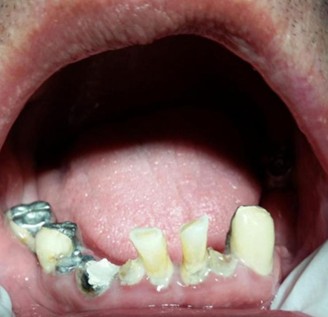
Figure 1: Periodontitis resulting from antineoplastic drugs (17).
Antineoplastic drugs affect bone marrow, leading to a reduction in thrombocyte production, which results in thrombocytopenia. The suppression of bone marrow can cause excessive bleeding, as well as the development of petechiae, ecchymoses, and hematomas. In the oral cavity, areas that are particularly vulnerable to excessive bleeding include the lower lip, the floor of the mouth, the soft palate, and the vestibular mucosa (29). Patients exhibiting a platelet count of less than 50,000/mm³ are generally considered contraindicated for undergoing tooth extractions due to the heightened risk of excessive bleeding. Specifically, individuals with platelet counts falling below 20,000/mm³ face a significant risk of hemorrhagic complications, which may present as gingivitis or more severe bleeding episodes. In the event of hemorrhage, clinical management typically involves the application of vasoconstrictors to facilitate hemostasis, with epinephrine being the most commonly employed agent due to its efficacy in reducing vascular permeability and promoting vasoconstriction (29).
Taste dysgeusia is a notable side effect of antineoplastic drugs. Approximately 39% of patients receiving these medications report experiencing taste dysgeusia. Among those with dysgeusia, around 38% report a salty taste, while 22% experience a sweet-sour taste. This condition leads to dysphagia (17). The changes in taste that the patients experience as a side effect of chemotherapy are attributed to damage of certain cranial nerves such as the seventh, ninth, and tenth cranial nerves, zinc deficiency that causes taste buds defect, vitamin D deficiency, or oral mucositis (30).
Effect of antineoplastic drugs on hard tissue
Antineoplastic drugs such as bisphosphonates cause osteonecrosis of the jaws by decreasing the blood supply to the jaws. Bisphosphonates are prescribed to treat malignant hypercalcemia, malignant melanoma, osteoporosis, and bone metastases (17). Osteonecrosis inhibits osteoclasts and osteoblasts’ function. Osteonecrosis can also result from trauma, biopsy, periodontal procedures, tooth extraction, or types of malignancies (31). Osteonecrosis typically presents initially in an asymptomatic manner; however, as the condition progresses, it manifests as pain accompanied by tooth mobility, ulceration, swelling, and paresthesia (31).
The diagnosis of osteonecrosis of the jaws depends primarily on the patient’s history and the presence of exposed bone (Figure 2) in the oral cavity for a period longer than eight weeks (32). Radiography does not help diagnose osteonecrosis of the jaws; however, a computed tomography (CT) Scan can delineate the extent of the disease (32).
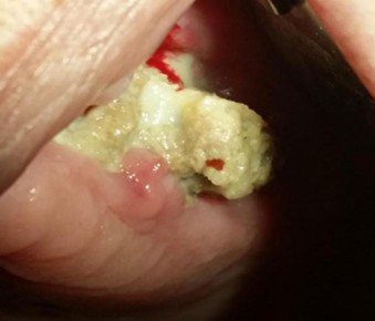
Figure 2: Osteonecrosis of the maxilla (17).
Treating osteonecrosis of the jaws includes oral hygiene protocol, antibiotic rinses, and systemic antibiotics. Non-responsive osteonecrosis requires ostectomy of the lesion and resection of the margins that extend into healthy areas (32).
Antineoplastic drugs affect the teeth as well. They cause microdontia, retardation of tooth development, enlargement of the pulp chamber, and root malformation (33). The extent of the disease depends on the age of the patient at the initiation of the chemotherapy and the number of cells affected (33). Furthermore, antineoplastic drugs cause enamel hypoplasia by damaging ameloblastic cells, hence causing enamel opacities (34). The administration of elevated doses of antineoplastic agents has been associated with significant adverse effects on dental development, specifically leading to root agenesis and tooth agenesis. This phenomenon occurs due to the disruption of Hertwig's epithelial root sheath, a critical structure in tooth development. The altered positioning of this sheath results in the malformation of the dental pulp chamber and affects the bi-furcation area of the roots. Consequently, the integrity of the dental architecture is compromised, potentially leading to a range of complications, including increased susceptibility to dental anomalies and delayed eruption, which may have long-term implications for oral health. (34). Additionally, antineoplastic drugs cause hypodontia by affecting tooth bud formation (33).
Conclusion
Antineoplastic agents are fundamental in the management of various malignancies, yet their therapeutic benefits are often accompanied by significant adverse effects that can compromise patient health. Systemic toxicities, such as myelotoxicity, neutropenia, thrombocytopenia, and increased susceptibility to infections and sepsis, can occur as a result of these medications. Additionally, these drugs can have specific detrimental effects on the oral cavity, manifesting as oral mucositis, dysgeusia, osteonecrosis, and periodontitis. Despite the critical need for effective cancer treatment, there remains a notable deficiency in research elucidating the mechanisms underlying these adverse effects and the strategies to mitigate them, underscoring an area ripe for further investigation.
Disclosure
Conflict of interest
There is no conflict of interest.
Funding
No funding.
Ethical consideration
Non applicable.
Data availability
Data that support the findings of this study are embedded within the manuscript.
