Volume 5, Issue 2
February 2025
Dental Management of a Patient With VACTERL Association Under General Anesthesia: A Case Report
Anas Shabra, Mostafa Marzouk, Sarah Albalawi
DOI: http://dx.doi.org/10.52533/JOHS.2025.50201
Keywords: Pediatric dentistry, VACTERL association, Dental management, Congenital anomalies, Tracheostomy
Introduction: VACTERL association is a rare congenital multisystem malformation that includes vertebral anomalies, anal atresia, cardiac malformations, tracheoesophageal fistula with or without esophageal atresia, renal dysplasia, and limb abnormalities. Clinicians usually require at least 3 of the mentioned anomalies for diagnosis. Its prevalence has been estimated between 1/10,000 to 1/40,000 infants. Both genetic and environmental factors may contribute to this association. Oral health in these patients is frequently overlooked due to their medical condition. This report discusses the dental findings and management of VACTERL association patient under general anesthesia.
Case Description: A 7-year-old Saudi boy, a known case of VACTERL association with a tracheostomy tube due to bilateral vocal cord paralysis, hyperactive airway disease, and a chromosome 5 anomaly. According to American society of anesthesiologists (ASA), his condition is classified as ASA III. He presented to the pediatric dental clinic with his mother, complaining of retained lower primary anterior teeth. Intraoral examination revealed poor oral hygiene, indicating parental neglect. The patient also has multiple carious lesions and retained lower primary central incisors with permanent ones erupting lingually. Due to the patient’s uncooperative behavior and his medical complexity, dental rehabilitation under general anesthesia was planned. Multidisciplinary coordination ensured safe anesthesia management, including a tracheostomy tube exchange. Treatment involved restorations, sealants, and oral hygiene counseling. The procedure was completed without complications, and follow-up care was scheduled to maintain oral health.
Conclusion: Preventive dental care is extremely important in these types of cases. Dental rehabilitation under general anesthesia for patients with VACTERL association may be challenging and needs to be adequately planned with a multidisciplinary team.
Introduction
VACTERL association is a rare congenital multisystem malformation that includes vertebral anomalies, anal atresia, cardiac malformations, tracheoesophageal fistula with or without esophageal atresia, renal dysplasia, and limb abnormalities (1, 2). It was first termed VATER, without cardiac and limb malformations, by Quan and Smith in 1970 (3). It is an association rather than a syndrome because these anomalies occur together more frequently than expected by chance, and there is no evidence for a single cause. For diagnosis, many clinicians require the presence of at least three malformations without clinical or laboratory-based evidence of overlapping conditions (1).
The prevalence of the VACTERL association has been estimated between 1/10,000 to 1/40,000 infants (approximately <1–9/100,000 infants) (1). VACTERL association has a multifactorial etiology, meaning environmental triggers interact with genetic factors (1, 4). Positive family history and high rates among monozygotic twins suggest a genetic role in VACTERL association (4). Multiple environmental triggers have been reported, including maternal diabetes, infertility treatment, in utero exposure to estrogen and/or progesterone-containing compounds, statins, and lead (1).
Multiple surgeries are often required during childhood, and various physical sequelae may follow, such as scoliosis, bowel dysfunction, gastroesophageal reflux, dysphagia, airway morbidity, and decreased cardiac, renal, or limb function (1). Chronic conditions seem to have a negative impact on psychosocial health, academic functioning, and social competence in children (5,6). Children with chronic conditions frequently undergo multiple hospital stays, endure painful medical procedures, face interruptions in their school attendance, and encounter restrictions on their social and physical activities, all of which can diminish their psychosocial well-being (7).
Being a parent of a child with VACTERL association can be taxing, with mixed emotional reactions, and shared responsibility for the child’s treatment and care. A child’s complex malformation often entails long-term follow-up, repeated episodes of anesthesia and procedures, and difficulties in everyday functions. Psychological processing, good medical care, and support from experts and other parents are essential in the parents’ struggle to reach self-confidence and adaptation. A care plan with individualized tailored care for each child including a training and support plan for the parents is warranted. An example would be to reduce the discrepancies in knowledge and experiences described between the local and tertiary hospital, by providing video sessions with the parents and responsible professionals at the local and tertiary hospital. This could be an appropriate mode of transferring information at discharge and during the follow-up of the child (8).
VACTERL syndrome poses risks in terms of anesthesia due to the presence of multiple anomalies. The risk of aspiration increases in VACTERL patients due to regurgitation and tracheoesophageal fistula (9). VACTERL children may have other associated abnormalities, such as cleft lip and palate, hemifacial microsomia (hypoplastic mandible with or without webbed neck), and may undergo different procedures. A difficult airway and intubation should be anticipated and demand careful pre-operative assessment and the use of skillful anesthetic techniques to avoid fatal complications (10).
Due to other critical malformations that may be life-threatening in some conditions, little concern is given to the oral health of those with VACTERL association. A few oral/dental findings of the VACTERL association have been reported in previous studies, including fusion (11), hypodontia (12), supernumerary teeth, and early eruption of permanent teeth (13).
This study aims to report a comprehensive overview of the dental findings and the multidisciplinary approach in managing patients with VACTERL association under general anesthesia.
Case Presentation
A 7-year-old boy who is a known case of VACTERL association presented to the dental medical compromised clinic at King Abdulaziz Medical City Hospital, National Guard, Jeddah. The mother was concerned about the lingual eruption of the lower permanent central incisors. This was the first dental visit for the patient. He was a full-term normal delivery to consanguineous parents. He is the only child, with no known family history of any congenital or hematological diseases. He was admitted to NICU for 4 months due to tracheoesophageal fistula with esophageal atresia and gastroesophageal reflux disease (GERD). He has bilateral vocal cord paralysis, and a tracheostomy was performed at one month of age. He has a single right kidney with a normal anatomy. Multiple hospital admissions and surgeries were done, including regular esophageal dilatation and bilateral laser cordectomy of the vocal cords. Chromosomal analysis of the patient showed 46XY with deletion of chromosome 5. On presentation, the patient weighs 14.5 kg and is 111 cm tall, which indicates he is underweight based on the National Center for Chronic Disease Prevention and Health Promotion criteria (2000). This delay in development is a result of his condition.
Extraorally, he exhibited a convex profile and normal facial appearance with competent lips. Intraoral examination revealed poor oral hygiene, indicating the parents neglected brushing the child’s teeth and did not care about the importance of oral hygiene, given his medical condition. The oral mucosa, frenulum, and tongue were within normal limits. Generalized physiological brown discoloration was present on the gingiva. He had carious right primary maxillary central incisor (#51) and first molar (#54), carious right mandibular second molar (#85), and retained lower primary central incisors with permanent ones erupting lingually (Figure 1).
Based on American association of pediatric dentistry (AAPD) guidelines, an orthopantomograph, right and left bitewing, and selected periapical x-rays were taken, which showed multiple interproximal caries in primary molars (Figure 2). Due to the uncooperative behavior and medical condition of the patient, dental rehabilitation under general anesthesia was the best option. The parents approved the treatment plan and signed the consent form.
Consultation with his primary physician and anesthesiologist about his ability to tolerate dental treatment under general anesthesia was conducted. The patient was cleared, classifying him as ASA III according to the American Society of Anesthesiologists (ASA) Physical Status Classification. The procedure preparation was done in cooperation with ear, nose and throat (ENT), respiratory therapy, and anesthesia teams.
On the day of the dental procedure, anesthesia was started after the ENT team was on board to change the uncuffed tracheostomy tube to a cuffed one to avoid significant leaks that could compromise mechanical ventilation. After preoxygenation, anesthesia started with Propofol 40 mg slowly IV, maintaining the patient’s spontaneous breathing to gain time during the tracheostomy tube exchange, as difficulty was expected. The leak was tested and was acceptable, not compromising mechanical ventilation. A throat pack was applied, the patient was connected to the ventilator, and anesthesia was maintained with inhalational Sevoflurane 2. Ventilation maintained on pressure-controlled ventilation-volume guaranteed (PCV-VG) mode. Dexamethasone 1.5 mg and Paracetamol 450 mg were given. The procedure was completed safely without complications. The throat pack was removed at the end of the procedure and the patient regained consciousness and recovered. In the Post Anesthesia Care Unit (PACU), the tracheostomy tube changed back to its original uncuffed size 4 by the ENT team after ensuring that the patient was fully awake and vitally stable.
Teeth (#55,54,64,75,74,84) were restored with stainless steel crowns, and right mandibular second molar (#85) was restored with a composite filling. All permanent first molars were sealed with resin-based sealants (Figure 3 and 4). The patient was put conservative program, which included dietary suggestions, oral hygiene motivation, fluoride application, and a maintenance program for regular follow-ups.
After being closely observed, the patient was discharged with a stable condition on the same day of the procedure. The parents were motivated to schedule regular follow-ups beginning two weeks after the general anesthesia.

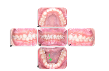
Figure 1: Intraoral examination shows early mixed dentition stage with multiple carious teeth and lingually erupted lower central incisors (green arrow).
Clinical intraoral preoperative photographs of the patient.
(A) Occlusal view of the maxillary arch showing mixed dentition and multiple carious lesions.
(B) Preoperative right lateral view of the dentition
(C) Preoperative frontal view of the dentition
(D) Preoperative left lateral view of the dentition
(E) Occlusal view of the mandibular arch with a green arrow pointing to lingually erupted lower central incisors.

Figure 2: Panoramic and bitewings radiographs show multiple carious teeth (red arrows); dental age coincides with chronological age.
Radiographic findings of the patient.
(A) Panoramic radiograph showing the overall dental and jaw structures. Shows dental age coincides with chronological age.
(B) Bitewing radiograph (left side) showing proximal caries in the primary molars (red arrows).
(C) Bitewing radiograph (right side) showing additional proximal carious lesions in the primary molars (red arrows).

Figure 3: Post-restorative treatment results under general anesthesia.
Postoperative intraoral photographs following dental rehabilitation.
(A) Occlusal view of the maxillary arch showing restored stainless-steel crowns on the primary molars.
(B) Right lateral view of the dentition.
(C) Frontal view of the dentition after extraction of primary upper central incisors.
(D) Left lateral view of the dentition.
(E) Occlusal view of the mandibular arch with stainless steel crowns on primary molars.

Figure 4: Post-radiographic treatment results under general anesthesia.
Postoperative Bitewings
(A) Postoperative right bitewing radiograph showing stainless steel crowns on the restored primary molars.
(B) Postoperative left bitewing radiograph displaying stainless steel crowns on the restored primary molars.
Discussion
VACTERL association can be diagnosed if at least three of the following malformations are observed: anal atresia, vertebral anomalies, cardiovascular defects, tracheoesophageal fistula, esophageal atresia, renal and/or radial anomalies, and/or limb anomalies. In our case, the patient had esophageal atresia, tracheoesophageal fistula, and renal anomalies. Additionally, the patient had GERD and bilateral vocal cord paralysis, which further complicated management.
As with many rare congenital diseases, VACTERL association requires a specific and multidisciplinary approach to treatment. Similar to other reported studies, this multidisciplinary approach proved successful. Due to the patient’s uncooperative behavior, dental treatment under general anesthesia was deemed necessary. Preoperative consultation and case preparation were conducted with the child’s primary physician, anesthesia team, respiratory therapy, and ENT teams to assess the patient’s ability to tolerate the procedure safely.
Extubating patients with difficult airways must be approached with considerable caution. Depending on the procedure, some children may require postoperative intensive care ventilation, while others may be safely extubated on the operating table. A history of conditions that increase aspiration risk, such as cleft lip/palate or tracheoesophageal fistula, may prevent early extubation. The pre-existing anomalies and potential risk of regurgitation in patients with VACTERL syndrome demand careful preoperative assessment and skilled anesthetic techniques to avoid fatal complications (10).
Additionally, anomalies such as fusion, hypodontia, supernumerary teeth, and early eruption of teeth may be influenced by other factors beyond VACTERL association, such as cleft lip and palate, hyperthyroidism, and preterm birth.
There are limited studies reporting dental findings in VACTERL association patients. One study found three missing permanent tooth buds (left upper lateral incisor and lower second premolars), while the upper right incisor was microdontic (11). However, in our case, no missing teeth were observed. Another study reported supernumerary teeth and early eruption of teeth (12), which were also absent in this case. It is worth noting that supernumerary teeth and early tooth eruption may be attributed to factors other than VACTERL association, such as preterm birth or hyperthyroidism. Teeth fusion has also been reported in one study (13), but this was not observed in our patient.
The patient presented with poor oral hygiene and significant tooth decay, aligning with other studies that report higher decay indices in patients with special healthcare needs. This increased decay may be attributed to additional pressures related to managing their medical conditions and hospitalization (14). Preventive dental care is extremely important in these types of cases, yet it is often overlooked as parents may feel overwhelmed by the broader medical challenges associated with VACTERL. This highlights the need for healthcare providers to educate families on the importance of regular dental care.
GERD was also a contributing factor in this case, as it increases acid production in the oral cavity, accelerating tooth erosion and loss. This aligns with studies reporting strong associations between GERD and tooth erosion (15-17). Gastric acid displaces saliva from tooth surfaces, while proteolytic pepsin removes protective dental pellicles, leading to selective dissolution of tooth components and eventual loss of substance (18, 19).
In this case, stainless steel crowns (SSCs) were chosen as the restorative treatment for posterior primary teeth. SSCs enhance the longevity of teeth and maintain oral function, consistent with findings from a systematic review demonstrating their excellent clinical performance as restoratives for posterior primary teeth (20).
This case aligns with previous reports on the challenges of managing patients with special healthcare needs but highlights the importance of tailoring dental treatment plans to individual medical complexities. The absence of dental anomalies, such as hypodontia or supernumerary teeth, contrasts with findings in other studies and suggests variability in VACTERL presentation.
To achieve the best possible outcomes, it is crucial to take a comprehensive medical history, communicate with the patient’s physician, guide the child, and reinforce good oral hygiene practices. Studies have shown that special healthcare needs patients with irregular dental checkup visits are approximately four times more likely to undergo repeated general anesthesia than those with regular checkups and adherence to oral hygiene instructions (14, 21).
This study is limited by its design a single case report, which prevents generalizing the dental findings and the management to all VACTERL association patients. Another limitation is the lack of long term follow up which prevents the evaluation of the outcomes of the dental management.
Dental rehabilitation under general anesthesia in patients with VACTERL association remains challenging and requires comprehensive case preparation. By fostering collaboration between medical and dental practitioners and emphasizing preventive care, improved outcomes can be achieved for this population. Future research should focus on long-term outcomes of restorative approaches, predictors of dental anomalies in VACTERL, and refining preventive and management strategies.
Conclusion
Managing dental conditions in patients with VACTERL association requires careful planning due to the complex interplay of systemic anomalies and associated risks, such as airway difficulties. This case highlights the importance of a multidisciplinary approach involving thorough preoperative assessment and collaboration among medical and dental teams to address both medical and dental complexities effectively.
Disclosure
Ethical committee approval
The ethical approval for this case report was obtained from the Institutional Review Board of King Abdullah International Medical Research Center with IRB No. IRB/2128/23. Written informed consent was acquired from the parents.
Competing interests
The authors declare that they have no competing financial, professional, or personal interests that might have influenced the performance or presentation of the work described in this case report.
Funding
The case report did not receive any funding from any sources.
Authors' contributions
AS has made substantial contribution to conceptualization, data curation, writing review & editing, visualization, supervision and project administration.
SB has a substantial contribution to investigation, data curation and writing the original draft.
All authors read and approved the final manuscript.
Acknowledgements
The authors would like to express their deep appreciation and thanks to the dental department and medical records staff for their cooperation and assistance during the case report preparation.