Volume 6, Issue 6
June 2026
Neuroimaging Patterns in Substance-Induced Psychotic Disorders
Doha Jamal Ahmad, Sara Ali Alhasan, Khadija Mohamed Aldallal, Mariam Hadi Alshammari, Abdullah Rshood AlRshood, Ali Hadi Alyami, Bader Hadi Alshammari, Faisal Mansour Alrowili
DOI: http://dx.doi.org/10.52533/JOHS.2026.60607
Keywords: substance-induced psychotic disorder, neuroimaging, magnetic resonance imaging, positron emission tomography, psychosis biomarkers
Substance-induced psychotic disorder (SIPD) is a clinically significant condition characterized by the emergence of psychotic symptoms following the use, intoxication, or withdrawal of psychoactive substances. The increasing prevalence of substance use worldwide, coupled with the growing recognition of psychosis as a severe neuropsychiatric consequence of substance exposure, has intensified interest in understanding the underlying neurobiological mechanisms of the disorder. Neuroimaging has become a valuable tool for investigating structural, functional, and neurochemical abnormalities that may contribute to symptom development. Evidence from magnetic resonance imaging (MRI), functional MRI, diffusion tensor imaging, positron emission tomography, and single-photon emission computed tomography has identified abnormalities in several brain regions involved in cognition, emotional regulation, salience attribution, and reward processing. Frequently implicated areas include the prefrontal cortex, hippocampus, amygdala, thalamus, and striatum. Structural imaging studies have demonstrated alterations in gray matter volume and white matter integrity, while functional imaging has revealed disruptions in neural connectivity and network organization. Molecular imaging findings have highlighted disturbances in dopaminergic and other neurotransmitter systems that parallel mechanisms observed in primary psychotic disorders. Neuroimaging data also suggest the presence of substance-specific patterns of brain dysfunction. Psychosis associated with methamphetamine use is commonly linked to frontostriatal abnormalities and dopaminergic dysregulation, whereas cannabis-related psychosis has been associated with hippocampal and cortical alterations. Despite considerable overlap with schizophrenia-spectrum disorders, distinct imaging profiles may exist that reflect the neurobiological effects of different substances. The identification of reliable neuroimaging biomarkers remains challenging due to methodological heterogeneity, variations in substance exposure, and the complex interaction between substance-related effects and underlying vulnerability to psychosis. Nevertheless, advances in multimodal imaging and computational analysis continue to improve our understanding of SIPD and may facilitate more accurate diagnosis, risk stratification, and treatment planning.
Introduction
Substance-induced psychotic disorder (SIPD) is a significant neuropsychiatric condition characterized by the emergence of hallucinations, delusions, and other psychotic symptoms temporally associated with the use, intoxication, or withdrawal of psychoactive substances. The disorder has gained increasing clinical and public health attention due to the rising prevalence of substance use worldwide and the growing recognition of psychosis as a severe consequence of exposure to certain substances, including cannabis, methamphetamine, cocaine, hallucinogens, and synthetic psychoactive compounds. Although psychotic symptoms associated with substance use were once considered transient and self-limiting, accumulating evidence suggests that a substantial proportion of affected individuals experience persistent symptoms or subsequently develop primary psychotic disorders, particularly schizophrenia-spectrum conditions (1)
The pathophysiology of SIPD is complex and multifactorial, involving interactions between genetic vulnerability, environmental factors, neurodevelopmental processes, and substance-related neurotoxicity. Psychoactive substances exert their effects through diverse neurotransmitter systems, most notably dopaminergic, glutamatergic, serotonergic, and endocannabinoid pathways. Dysregulation of these systems can disrupt normal brain function and contribute to the emergence of psychotic symptoms. Among these mechanisms, alterations in dopaminergic transmission have been extensively implicated, given their established role in both substance use disorders and psychotic illnesses (2). The neurobiological substrates underlying SIPD remain incompletely understood, creating challenges for accurate diagnosis, prognosis, and treatment.
Advances in neuroimaging techniques have provided valuable opportunities to investigate the neural correlates of psychosis associated with substance use. Structural magnetic resonance imaging (MRI), functional MRI, diffusion tensor imaging, positron emission tomography (PET), and single-photon emission computed tomography (SPECT) have facilitated the characterization of brain abnormalities in affected individuals. These modalities have revealed alterations in cortical and subcortical regions involved in cognition, reward processing, emotional regulation, and executive functioning. Brain regions commonly implicated include the prefrontal cortex, hippocampus, amygdala, striatum, and thalamus, many of which overlap with those reported in primary psychotic disorders (3).
Neuroimaging studies have also highlighted the heterogeneity of SIPD across different substances. Methamphetamine-associated psychosis has been linked to structural and functional abnormalities involving frontostriatal circuits, whereas cannabis-related psychosis has been associated with alterations in hippocampal and cortical networks. Similarly, cocaine-induced psychosis has demonstrated changes in reward-related and limbic pathways. These findings suggest that while common neurobiological mechanisms may contribute to psychosis across substances, substance-specific patterns of brain dysfunction may also exist (4).
Review
Neuroimaging findings in substance-induced psychotic disorders suggest substantial overlap with the neural abnormalities observed in primary psychotic disorders, particularly within frontostriatal, limbic, and thalamocortical networks. Structural imaging studies have consistently demonstrated alterations in gray matter volume and cortical integrity in regions associated with executive functioning, emotional regulation, and reward processing. These abnormalities may reflect the neurotoxic effects of prolonged substance exposure, although pre-existing vulnerabilities cannot be excluded. Functional imaging studies further indicate disruptions in connectivity and metabolic activity, supporting the hypothesis that substance use may precipitate psychosis through dysregulation of neural circuits already implicated in schizophrenia-spectrum disorders (5).
Despite these similarities, emerging evidence points toward the existence of substance-specific neuroimaging signatures. Methamphetamine-associated psychosis has frequently been linked to abnormalities within dopaminergic pathways and frontostriatal circuitry, whereas cannabis-related psychosis appears more strongly associated with hippocampal and cortical alterations. Such differences may explain variations in symptom presentation, disease course, and risk of transition to chronic psychotic disorders. Nevertheless, heterogeneity in imaging methodologies, sample characteristics, and patterns of substance use remains a significant limitation across studies. Future investigations employing multimodal imaging approaches and longitudinal designs may help clarify whether observed abnormalities represent transient substance-related effects or enduring biomarkers of psychosis vulnerability. Improved characterization of these patterns could enhance diagnostic accuracy and support the development of more targeted therapeutic strategies (6).
Structural and Functional Neuroimaging Abnormalities
Neuroimaging studies have provided important insights into the brain alterations associated with substance-induced psychotic disorder, revealing abnormalities that span both structural and functional domains. MRI investigations frequently identify changes in cortical and subcortical regions implicated in cognition, emotional regulation, salience attribution, and reward processing. Among individuals experiencing psychosis related to stimulant use, reductions in gray matter volume have been observed within the prefrontal cortex, anterior cingulate cortex, hippocampus, and temporal regions. These areas are closely linked to executive control, memory, and reality monitoring, functions that are often disrupted during psychotic episodes. Structural abnormalities have also been reported in the striatum and thalamus, supporting the involvement of circuits that modulate dopaminergic signaling and sensory information processing (7).
Diffusion-based imaging techniques have expanded understanding of white matter integrity in substance-induced psychosis. Alterations in major white matter tracts connecting frontal and limbic structures suggest impaired communication between regions responsible for cognitive control and emotional processing. Such findings are particularly evident in methamphetamine-associated psychosis, where prolonged exposure has been associated with microstructural abnormalities involving frontostriatal pathways. Disturbances within these networks may contribute to impaired decision-making, heightened salience attribution, and the persistence of psychotic symptoms even after cessation of substance use. Neuroanatomical changes observed in stimulant-related psychosis often resemble those described in schizophrenia, raising questions regarding shared neurobiological mechanisms and the possibility that substance exposure may accelerate pathological processes in vulnerable individuals (8).
Functional neuroimaging studies provide complementary evidence by demonstrating abnormalities in brain activity and connectivity. Functional MRI investigations have identified disrupted communication between cortical and subcortical networks, particularly involving the thalamus, prefrontal cortex, and default mode network. Altered thalamocortical connectivity has attracted considerable interest because similar patterns have been associated with psychotic symptom severity and disturbances in sensory integration. Experimental studies involving psychoactive substances capable of inducing psychosis-like experiences have reported connectivity changes that parallel those seen in clinical psychotic disorders, suggesting common neural pathways underlying aberrant perception and thought processes (9).
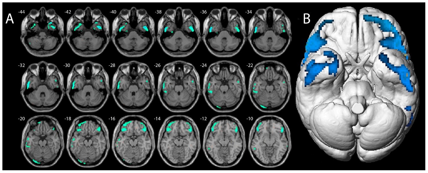
Studies that included PET and SPECT have further highlighted neurochemical and metabolic abnormalities associated with substance-induced psychosis. Functional neuroimaging findings frequently reveal regional metabolic disturbances that may not be evident on conventional structural imaging. Reduced cerebral glucose metabolism has been reported in cortical regions involved in executive functioning, attention, and reality monitoring. An example of these abnormalities is illustrated in Figure 1, which demonstrates significant hypometabolism in brain regions associated with psychotic symptomatology.

Figure 1: Axial magnetic resonance slices (A) and brain surface rendering (B) show the results of SPM analysis. The color-coded regions indicate the locations where the patients’ voxel values are significantly hypometabolic compared with the healthy control group (10).
Variations in glucose metabolism, dopamine receptor availability, and regional cerebral blood flow have been documented across different substance categories. Cannabis-related psychosis has been linked to alterations in dopaminergic function and limbic activity, while stimulant-induced psychosis frequently demonstrates metabolic disturbances involving frontal and temporal regions. The integration of structural MRI with PET imaging has revealed concurrent morphological and metabolic alterations, indicating that anatomical changes are often accompanied by functional dysregulation across interconnected neural systems involved in psychosis expression (11).
Clinical and Pharmacological Significance of Neuroimaging Findings
Psychotic symptoms arising in the context of substance use often resemble those observed in primary psychotic disorders, creating uncertainty regarding diagnosis, prognosis, and treatment selection. Structural and functional imaging findings offer valuable information regarding the neural substrates involved in symptom generation and may assist in identifying biological features that distinguish transient substance-related psychosis from conditions associated with a higher likelihood of chronic psychotic illness. Neuroimaging abnormalities involving prefrontal, temporal, and limbic regions have been linked to cognitive impairment, affective dysregulation, and persistent psychotic symptoms, suggesting that imaging markers may contribute to risk stratification in clinical settings (12).
Functional imaging studies have also enhanced understanding of the neurochemical mechanisms underlying psychosis associated with psychoactive substances. Alterations in dopaminergic activity remain among the most consistently reported findings, particularly in relation to cannabis and stimulant exposure. Molecular imaging investigations have demonstrated changes in dopamine receptor availability and neurotransmitter signaling that parallel mechanisms implicated in schizophrenia. These observations support the rationale for using antipsychotic medications that target dopaminergic pathways, while also highlighting the possibility that pharmacological responses may vary according to the specific neural alterations induced by different substances. Imaging-derived evidence therefore provides a biological framework for understanding why certain individuals experience symptom resolution following abstinence, whereas others require prolonged pharmacological intervention (13).
Neuroimaging has also contributed to the evaluation of treatment-related brain changes. Longitudinal studies examining pharmacological interventions in psychotic disorders have identified modifications in brain structure, connectivity, and neural activity following antipsychotic treatment. Such findings indicate that therapeutic effects extend beyond symptom reduction and may involve measurable alterations within neural circuits associated with psychosis. In the context of substance-induced psychotic disorder, these observations are particularly relevant because they suggest potential imaging markers for monitoring treatment response and disease progression. The ability to detect neurobiological changes during recovery may facilitate more individualized treatment approaches and support clinical decision-making regarding the duration and intensity of pharmacotherapy (14).
Interest has also emerged in advanced imaging modalities capable of assessing medication-related effects directly within the brain. Pharmacological MRI and related techniques allow investigators to examine how specific drugs influence neural activity and connectivity patterns. These approaches have demonstrated potential utility in identifying biomarkers associated with treatment efficacy, adverse effects, and target engagement. As imaging technology continues to evolve, integration of radiological findings with clinical and pharmacological data may provide increasingly refined tools for managing psychosis occurring in the setting of substance use and for characterizing biologically distinct patient subgroups (15).
Neuroimaging Biomarkers and Diagnostic Challenges
The identification of reliable neuroimaging biomarkers for substance-induced psychotic disorder remains a major objective in psychiatric research. Diagnostic uncertainty is common because psychotic symptoms related to substance exposure frequently overlap with those observed in schizophrenia-spectrum and affective psychotic disorders. Hallucinations, delusions, disorganized thought processes, and cognitive dysfunction may present with comparable clinical features regardless of etiology, making it difficult to determine whether psychosis is primarily substance-induced or reflects the early manifestation of an underlying psychiatric condition. Neuroimaging has therefore attracted considerable interest as a potential source of objective biological markers capable of supporting diagnostic decision-making and improving prognostic accuracy (16).
Structural neuroimaging studies have demonstrated abnormalities involving cortical thickness, gray matter volume, and subcortical morphology in individuals with psychosis associated with substance use. Findings from first-episode psychosis research indicate that imaging abnormalities are often detectable during the early stages of illness, although the specificity of these changes remains limited. Similar alterations have been described across multiple psychotic disorders, including substance-related psychosis, reducing their utility as standalone diagnostic markers. Variability in the type of substance used, duration of exposure, age of onset, and periods of abstinence further complicates interpretation of imaging findings. Distinguishing neurotoxic effects of substances from pre-existing neurodevelopmental vulnerabilities remains a persistent challenge in clinical neuroimaging research (17).
Functional and molecular imaging approaches offer additional opportunities for biomarker discovery by characterizing abnormalities in neurotransmitter systems and neural network activity. Positron emission tomography studies have highlighted alterations in dopaminergic signaling, glutamatergic pathways, and neuroinflammatory processes associated with psychosis. Such findings are particularly relevant because they correspond closely with established neurobiological models of psychotic disorders. Molecular imaging has also provided insight into early psychosis-related neurochemical changes, suggesting that biomarker development may benefit from integrating structural, functional, and neurochemical data rather than relying on a single imaging modality. Multimodal approaches may improve sensitivity to clinically meaningful differences between substance-induced psychosis and primary psychotic illnesses (18).
Interest has increasingly shifted toward advanced biomarker frameworks that combine neuroimaging findings with clinical, cognitive, genetic, and pharmacological data. Contemporary psychiatric biomarker research emphasizes network-level dysfunction rather than isolated regional abnormalities, reflecting the complex and distributed nature of psychosis-related brain changes. Emerging evidence suggests that machine learning and computational neuroimaging techniques may help identify distinct biological profiles among individuals presenting with psychotic symptoms. These developments hold promise for enhancing diagnostic precision, particularly in cases where substance use obscures the clinical picture and conventional diagnostic criteria provide limited certainty regarding the underlying nature of the disorder (2).
Conclusion
Neuroimaging studies have revealed consistent structural, functional, and neurochemical abnormalities associated with substance-induced psychotic disorder, highlighting the involvement of brain networks implicated in cognition, reward processing, and psychosis. Current evidence suggests both shared and substance-specific neurobiological patterns that may influence clinical presentation and outcomes. The integration of multimodal imaging approaches has expanded understanding of disease mechanisms and potential biomarkers relevant to diagnosis and treatment. Continued advances in neuroimaging research may support more precise differentiation of substance-induced psychosis from primary psychotic disorders and contribute to personalized clinical management.
Disclosure
Conflict of interest
There is no conflict of interest.
Funding
No funding.
Ethical consideration
No considerations.
Data availability
All data is available within the manuscript.
Author contribution
All authors contributed to conceptualizing, data drafting, collection and final writing of the manuscript.